Pages

Thursday, June 4, 2015

Antibiotic alternatives rev up bacterial arms race


Sara Reardon - 27 May 2015


From predatory microbes to toxic metals, nature is inspiring new ways to treat infections.


More than eight decades have passed since Alexander Fleming’s discovery of a fungus that produced penicillin — a breakthrough that ultimately spawned today’s multibillion-dollar antibiotics industry. Researchers are now looking to nature with renewed vigour for other ways of fighting infection.

Few new antibiotics are in development, and overuse of existing ones has created resistant strains of deadly bacteria. “We need a change from what we have,” says Stephen Baker, head of medicinal chemistry for antibacterials at Glaxo­SmithKline in College­ville, Pennsylvania.

Baker will talk about some of the alternatives to antibiotics on 2 June at the American Society for Microbiology’s annual meeting in New Orleans, Louisiana. Here are a few of the therapies that scientists are exploring.


Predatory bacteria


Bacteria cause infection, but some can also fight it by preying on fellow microbes. Several researchers are beginning to test these predatory bacteria in animal models and cell cultures.

The best-known species, Bdellovibrio bacteriovorus, is found in soil. It attacks prey bacteria by embedding itself between the host’s inner and outer cell membranes, and begins to grow filaments and replicate. “It’s like going into a restaurant, locking the door and starting to munch away,” says Daniel Kadouri, a bacteriologist at Rutgers University in Newark, New Jersey. The host bacterium eventually explodes and releases more B. bacteriovorus into the environment.

Kadouri and others are also studying the therapeutic potential of the predatory bacterium Micavibrio aeruginosavorus. And a team has engineered the gut bacterium Escherichia coli to produce peptides that kill Pseudomonas aeruginosa, a microbe that causes pneumonia.

This preliminary research is attracting attention. The Pathogen Predators programme of the US Defense Advanced Research Projects Agency, which aims to treat soldiers who contract infections on the battlefield, announced nearly US$16 million in research grants this week to groups studying predatory bacteria.


Antimicrobial peptides


Plants, animals and fungi have vastly different immune systems, but all make peptides — small proteins — that destroy bacteria. Peptides from creatures such as amphibians and reptiles, which are unusually resistant to infection, could yield new therapeutics.

Peptides with antibacterial activity have been isolated from frogs, alligators and cobras, among others, and some seem to be effective in epithelial cell cultures and at healing wounds in mice. These peptides can be modified to increase their potency, and several are in clinical trials. One, called pexiganan, based on a peptide from frog skin, is now in phase III clinical trials to treat diabetic foot ulcers.

But synthesizing such molecules can be expensive, a hurdle that scientists must overcome to bring new peptide drugs to market.


Phages


Of all the alternatives to antibiotics, phages — viruses that attack bacteria — have been used the longest in the clinic. Scientists in the Soviet Union began developing phage therapies in the 1920s, and former Soviet countries continue the tradition.

Phages have several advantages over antibiotics. Each type attacks only one type of bacterium, so treatments leave harmless (or beneficial) bacteria unscathed. And because phages are abundant in nature, researchers have ready replacements for any therapeutic strain that bacteria evolve to resist.

Mzia Kutateladze, who heads the scientific council at the Eliava Institute in Tblisi, Georgia, says that antibiotic resistance is driving more Western patients to phage-therapy clinics in Eastern Europe. The US National Institute of Allergy and Infectious Diseases in Bethesda, Maryland, now lists phages as a research priority for addressing the antibiotic crisis. A clinical trial of a phage treatment for infections associated with burns is planned by a consortium of European centres to start this summer.


Gene-editing enzymes


CRISPR, a gene-editing technique that has taken the scientific world by storm, is based on a strategy that many bacteria use to protect themselves against phages. Researchers are turning that system back on itself to make bacteria kill themselves.

Normally, the bacteria detect and destroy invaders such as phages by generating a short RNA sequence that matches a specific genetic sequence in the foreign body. This RNA snippet guides an enzyme called Cas9 to kill the invader by cutting its DNA.

Scientists are now designing CRISPR sequences that target genomes of specific bacteria, and some are aiming their CRISPR kill switches at the bacterial genes that confer antibiotic resistance.


Metals


Metals such as copper and silver are the oldest antimicrobials. They were favoured by Hippocrates in the fourth-century bc as a treatment for wounds, and were used even earlier by ancient Persian kings to disinfect food and water. Only now are researchers beginning to understand how metals kill bacteria.

Some groups are exploring the use of metal nanoparticles as antimicrobial treatments, although little research has been done in people. Because metals accumulate in the body and can be highly toxic, their use may be restricted mostly to topical ointments for skin infections.

An exception is gallium, which is toxic to bacteria that mistake it for iron, but is safe enough in people to be tested as an intravenous treatment for lung infections. This summer, researchers at the University of Washington in Seattle will begin a phase II clinical trial of gallium in 120 patients with cystic fibrosis. Pilot studies found that the metal was moderately successful at breaking down microbial biofilms in the lungs and improving patients’ breathing.


Nature 521, 402–403 (28 May 2015) doi:10.1038/521402a

Thursday, May 28, 2015

DNA Gains New Bases, Keeps Shapely Helix


Scientists have created a double helix out of six nucleotides,
 two more than what nature had devised.
[American Chemical Society]
Additions to DNA’s four-letter alphabet have been announced before, but the new letters don’t always fit comfortably into DNA’s double helix. Even slightly awkward letters can distort DNA’s overall shape and alter the molecule’s plasticity. Worse, a sequence of awkward letters that omits natural letters can amplify any structural problems, limiting new-letter-enriched synthetic DNA’s ability to interact with structural proteins and enzymes, jamming life’s molecular machinery.
A new pair of letters, however, seems to slide into DNA fairly easily. The new letters, or nucleotide bases, are called Z and P. They emulate structural and functional features of DNA’s natural nucleotide bases, C and G (cytosine and guanine) and A and T (adenine and thymine). Not only can they be interspersed with the natural nucleotide bases, they can be run adjacent to each other in a sequence, and the resulting DNA will still form a nice helix. What’s more,
the resulting DNA even shows the ability to evolve.
Z stands for 6-amino-5-nitro-2(1H)-pyridone. P stands for 2-amino-imidazo[1,2-a]-1,3,5-triazin-4(8H)one. Z and P were introduced in a pair of papers prepared by Millie M. Georgiadis, Steven A. Benner, and colleagues from Indiana and Florida, and published in the Journal of the American Chemical Society. (Dr. Georgiadis is affiliated with Indiana University− Purdue University Indianapolis, and Dr. Benner is affiliated with the Foundation for Applied Molecular Evolution, and Firebird Biomolecular Sciences.)
The first paper, “Structural Basis for a Six Nucleotide Genetic Alphabet”—appeared on May 11. It presented crystal structures to demonstrate that the new nucleotides paired with “geometries that are similar to those displayed by standard duplex DNA.” The second paper—“Evolution of Functional Six-Nucleotide DNA”—appeared a day later. It presented results form a laboratory in vitro evolution (LIVE) experiment to show that the new nucleotide bases could be incorporated into newly evolved, selectively binding species.
The first paper emphasized three important findings:
  1. The discovery that canonical Watson–Crick pairing survives in duplexes that contain multiple and multiple adjacent Z:P pairs.
  2. ZP-containing oligonucleotides adopt canonical helical forms, B- and A-form DNA. The ability of DNA to adopt A-form enables a number of important protein–DNA interactions such as those in the polymerase active site, which requires that the nucleobase pair within the active site and the adjacent pair adopt A-form in order to appropriately position the template-primer for optimal interactions with the polymerase increasing fidelity.
  3. The Z-nitro group imparts new properties to the major groove of DNA that can potentially be exploited for recognition by proteins.
The second paper explained how the LIVE experiment involved developing a GACTZP library, which was challenged to deliver molecules that bind selectively to liver cancer cells, but not to untransformed liver cells.
“Unlike in classical in vitro selection, low levels of mutation allow this system to evolve to create binding molecules not necessarily present in the original library,” the article’s authors wrote. “Over a dozen binding species were recovered. The best had Z and/or P in their sequences. Several had multiple, nearby, and adjacent Zs and Ps. Only the weaker binders contained no Z or P at all. This suggests that this system explored much of the sequence space available to this genetic system and that GACTZP libraries are richer reservoirs of functionality than standard libraries.”

Friday, May 8, 2015

Could CRISPR Be the Magic Bullet?


You Can Be “On Target” and Still Fail to Win a Prize

MaryAnn Labant


The research community’s rapid acceptance of the CRISPR/Cas technology is propelling a stage of deep investment in technology development. Already, three companies have emerged focusing on CRISPR therapeutic applications: Intellia Therapeutics, Editas Medicine, and CRISPR Therapeutics.

To continue to move the technology forward, scientists recently converged at the CRISPR Precision Gene Editing Congress to discuss unmet needs and new findings. The event, which took place in Boston, devoted particular attention to overcoming specificity, efficiency, and delivery challenges associated with the CRISPR/Cas9 system.

Many of these challenges relate to the mechanisms a cell may use to repair CRISPR/Cas-induced double-strand breaks (DSBs). A cell has two pathway choices. Non-homologous end joining (NHEJ), an error-prone ligation process, can result in small insertions and deletions (indels) at cleavage sites, whereas homology-directed repair (HDR) employs homologous DNA sequences as templates to make specific changes for precise repair. In most cells, NHEJ performs the majority of repair events.

Identifying and minimizing off-target events are major challenges. To meet these challenges, the Alt laboratory at Boston Children’s Hospital developed high-throughput genome translocation sequencing (HTGTS), an enzyme- and target-agnostic technique to rapidly expose potential off-target problems. Frederick W. Alt, Ph.D., and colleagues recently described the technique in an article that appeared in Nature Biotechnology.

“The method robustly detects DNA DSBs generated by engineered nucleases across the human genome based on their translocation to other endogenous or ectopic DSBs,” the article read. “HTGTS with different Cas9:sgRNA or TALEN nucleases revealed off-target hotspot numbers for given nucleases that ranged from a few or none to dozens or more, and extended the number of known off-targets for certain previously characterized nucleases more than 10-fold.”


When HTGTS was used to compare Cas9 nuclease and Cas9 paired nickases, paired nickases showed reduced off-target activity. Paired nickases were also assessed by scientists at Sigma-Aldrich.

“We compared paired nickases to Cas9-FokI nucleases. Paired nickases have about a 10-fold increase in design density, the number of nucleases that target a specific sequence in the selected area,” commented Gregory Davis, R&D manager, molecular biotechnology. “The more nuclease options, the better the chances of finding an active one near site-restricted locations such as disease single-nucleotide polymorphisms (SNPs).”

Like other companies, Sigma-Aldrich is evaluating methods to boost homologous recombination (HR) rates and inhibit NHEJ. Small molecules are being investigated, along with components of the DNA repair machinery such as mRNAs for RAD proteins. Enhancement techniques offer some improvement, but those improvements are not universally applicable to all cell types.

The company recently introduced a nuclease-based kinase knockout lentiviral library, but the challenge is increasing library screening effectiveness for cancer cell lines, which typically demonstrate some level of polyploidy. When the Cas9 nuclease library on the A459 lung cancer cell line was evaluated, a target diploid gene responded with a robust knockout, yet an expected knockout response for another gene was not seen. That particular gene turned out to be tetraploid.

Epigenetically based activators and inhibitors may be another approach, and the company is considering CRISPR-based gene regulation for inhibition or activation, CRISPRi or CRISPRa. Gene regulation may better simulate drugs that suppress activity and prove more effective than the nuclease-knockout method in lentiviral screening applications.


Measurement Systems

HDR and NHEJ editing events generally occur at low frequencies, necessitating ultrasensitive techniques for detection and quantification of edited alleles. While some studies have relied on NGS, a next-generation PCR technology called droplet digital PCR (ddPCR) is providing researchers with rapid, low-cost, ultrasensitive quantification of both NHEJ and HDR editing events.


ddPCR has already been widely used for high-sensitivity and high-precision applications such as rare cancer mutation detection and copy number analysis, noted Jennifer Berman, Ph.D., staff scientist, Digital Biology Center, Bio-Rad Laboratories.


Since HDR and NHEJ editing events can occur at very low frequency (<1%), especially HDR in primary or induced pluripotent stem (iPS) cells, ddPCR appears to be a fit for researchers wanting a rapid, sensitive, quantitative readout of editing in cells and tissues. The technique also enables empirical validation of guide RNA efficiency and measurement of the ratio of HDR:NHEJ at a targeted locus.


“ddPCR is one of the first sophisticated measurement systems for genome editing. The other option is sequencing, which is time-consuming, expensive and out of reach for most people,” explained Bruce Conklin, M.D., a senior investigator at the Gladstone Institute of Cardiovascular Disease and a professor of medicine at the University of California, San Francisco.


The Conklin laboratory works with iPS cells and is primarily interested in HDR, which is typically less than 1% of total alleles. A recent Nature Methods article by the group was the first demonstration that the genome could be changed one base at a time without any mark of an antibiotic reselection marker, a scarless replacement. Populations of cells that have a very rare cell with a single-base change are isolated using ddPCR as a measurement tool, then enriched sequentially, until a pure clone results, in a method termed sib-selection.


“With our method, you can see if the mutation you want is there from the start,” asserted Dr. Conklin. “Single base changes cause many human genetic diseases. To figure out the problem, you want to be able to change one thing and see what happens.”


“We are also looking at ddPCR to quantify HDR and NHEJ simultaneously to isolate conditions where there is more HDR than NHEJ,” he added. “Conditions are different in every cell type, for each location, and we do not understand the rules.”


Application to Animal Model

Mouse models have the potential to quickly screen and build a causal relationship between sequence variations in humans and their phenotypes. Historically, either pronuclear injection of a transgene into a mouse embryo or conventional gene targeting using embryonic stem (ES) cells produced new models.

In 2013, a study led by Rudolph Jaenisch, M.D., a professor of biology at MIT and a founding member of the Whitehead Institute for Biomedical Research, culminated in a published work that was the first to describe a CRISPR/Cas-engineered animal species. CRISPR’s ability to engineer targeted mutagenesis in the genome directly on the zygotes circumvents the need for germline-competent ES cells, and appears to result in more predictable models in a fraction of the previous time and cost.

The simplest way to create a knockout model is to inject CRISPR/Cas reagents, including Cas9 mRNA and a single gRNA, into the mouse embryo. If a knockin alteration is small, the intended mutation can be accommodated into a donor oligonucleotide of the maximal size of 200 bps; for larger alterations that cannot be engineered into a donor oligonucleotide, such as incorporation of a reporter gene or a human sequence, a donor plasmid is often used.

“We are now exploring the use of CRISPR for larger-scale genetic manipulation and humanization of the mouse genome. We do not know yet the size limitation of the genetic manipulation that you can introduce with the CRISPR/Cas technology,” discussed Wenning Qin, Ph.D., associate director of genetic engineering technologies, The Jackson Laboratory.

The Jackson Laboratory uses insertion of a fluorescent reporter gene into the Nanog locus, a gene expressed in early embryos, as the platform for parameter optimization. To determine if there were any off-target effects in addition to the on-target insertion of the reporter gene into the Nanog locus, two HDR mice carrying the reporter gene were genome sequenced, and evaluated minimally for the top 5,000 sites. No off-target effects were observed indicating that the CRISPR/Cas reagent used had a clean off-target profile among the examined sites.

Various means of enhancing the on-target efficiency of CRISPR/Cas9 modifications are being investigated by Taconic Biosciences. For example, the company is investigating the implementation of Cas9-orthologs to gain more flexibility in the choice of target sequences. It is also evaluating alternative delivery methods as well as experimental design and execution optimization.

To date, analyses have focused on evaluating on-target modifications. Nonetheless, various approaches to enhance specificity—for example, the use of Cas9 nickase, truncated sgRNAs, and Cas9 protein instead of mRNA—are being assessed. According to Jochen Welcker, Ph.D., senior manager of scientific development, improving the generation of more challenging types of alleles is a major development objective, and efficiency issues for specific applications need to be resolved.

For the generation of conditional knockout alleles, one obstacle is the efficient and correct integration of loxP-sites. Using ssODNs (single-stranded oligodeoxynucleotides) as donor material, for example, frequently leads to incomplete integration of the loxP-sites encoded by these ssODNs. The high efficiency of NHEJ-mediated deletion of the genomic region between the two loxP-sites is also a major hurdle as it leads to knockout alleles in up to 30% of the injected zygotes. The generation of complex knockin alleles is limited by the frequency of insertion/replacement of longer sequences by HDR.

Taconic has generated more than 200 humanized models by HDR. A drawback of this approach is the fact that only a single allelic variant can be modeled, while ever increasing amounts of human gene variants are being identified by genome-wide association studies (GWAS). Such variants can now be introduced quickly and efficiently, directly on existing humanized backgrounds by the use of CRISPR/Cas9 genome editing, making it possible to generate a whole range of human allelic variants from a single humanized-mouse model.

Bioinformatics Needs

“Bioinformatics plays an important role in CRISPR advancement. No-charge informatics packages are available, such as eCRISP, MIT tool, Doench activity scoring, and Zifit, that tackle a portion of the informatics aspects to CRISPR design, and many commercial companies have simple design tools built into their reagent-ordering systems,” stated Eric Rhodes, chief technology officer, Horizon Discovery. “Our new tool, gUIDEbook, a collaboration between Horizon and DesktopGenetics, is the first free application to combine all three aspects of informed design.”

First, all available protospacer adjacent motif (PAM) sites in a given region, which represent a potential gRNA design, must be found. Virtually all CRISPR design tools enable this and differ primarily in how the sequence is entered and user interface complexity. All programs essentially return the same information.

Second, off-target cutting potential by any given gRNA must be determined. Historically, searches found closely related sequences and scores were generated using weighting based on the number of mismatches, their location in the gRNA, and the number of occurrences in the genome. A new finding has demonstrated that “bulges” can occur in the matching of a gRNA to a potential target. More intensive searching is now required to identify all putative binding sites. The unknown is how likely an off-target identified by either method is actually going to be engaged.

The Doench algorithm, which is still early in its development, focuses solely on how much cutting activity a given gRNA design is likely to have. The algorithm calculates predictive scores on the basis of known guides and cutting activities. It does not account for off-target potential, so it has somewhat limited standalone value.

Delivery

Thermo Fisher Scientific supplies a complete workflow for gene editing and cell engineering that focuses on design, delivery, and analysis. Transfection-grade Cas9 protein and mRNA have been functionally tested in several cell lines, including iPS and ES cells, and both contain a nuclear localization signal (NLS) to aid in delivery. The GeneArt Cas9 Nuclease is extensively purified and quality controlled to remove nonspecific endonucleases and endotoxins.

According to Jason Potter, senior scientist of protein engineering, cell lines vary in how easily they can be transfected. With plasmids and mRNA, the cell must still process the transcripts and make Cas9 complexes before it can act. To simplify the process, the gRNA can be made and complexed with the transfection-grade Cas9 protein in vitro. After it is delivered by lipids or electroporation is used, the Cas9 complex is able to act once it reaches the nucleus. Analysis of the edited cells can then be done using the GeneArt Genomic Cleavage Detection kit or by sequencing.

Lipids, including Lipofectamine 3000, Lipofectamine RNAiMAX, and Lipofectamine MessengerMAX, have been used for delivery of plasmids and RNAs for years. Drawing on this knowledge of Cas9, the company has optimized lipid dosages and protocols for high transfection efficiency and low toxicity. Due to the exposed guide RNA component of the Cas9 complex, RNAiMAX also works for delivery of Cas9 protein. For electroporation, the key consideration is optimizing the voltage and pulse conditions for the cell line.

Gene Editing with Cas9 and Optimal Promoter

The Cas9 (CRISPR associated protein 9) system has gained significant interest due to its relative simplicity and ease of use compared to other genome-engineering technologies, according to many scientists. The CRISPR/Cas9 system requires a complex of the Cas9 protein with a trans-activating RNA (tracrRNA) and a gene-targeting CRISPR RNA (crRNA) or a single guide RNA (sgRNA, a chimeric form of tracrRNA with a crRNA).

Researchers at Dharmacon, now part of GE Healthcare, recently carried out a study on the efficiency of using synthetic crRNA and tracrRNA to introduce gene-editing events when co-transfected with a plasmid expressing Cas9. They explored the use of antibiotic and FACS methods for enrichment of cells that have undergone gene editing, and the use of multiple promoters to increase efficiency of gene editing with Cas9 and synthetic tracrRNA and crRNA.

The researchers reported that utilizing a highly active promoter for Cas9 expression enables better editing in specific cell lines. Enrichment of transiently transfected cells either by fluorescence-activated cell soring or puromycin selection can further improve the yield of edited cells, they added.

In addition, they concluded that efficient gene editing can be achieved with a three-component system: plasmid Cas9 and synthetic tracrRNA and crRNA. They also pointed out that use of synthetic tracrRNA and crRNA is a simplified method for gene editing of one or more genes without requiring any cloning steps, and that the three-component CRISPR/Cas9 system is amenable to high-throughput genome editing applications.

Thursday, May 7, 2015

New Stem Cell Identified


By Anna Azvolinsky | May 6, 2015

Researchers isolate an easy-to-manipulate, stable, and spatially distinct pluripotent cell type.


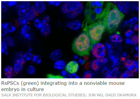
Scientists have isolated and defined a new type of pluripotent cell from early mouse embryos and from monkey and human stem cell lines. The monkey- and human-derived versions of these pluripotent cells can divide and generate the three germ layers in a developing mouse embryo, providing the first demonstration that human pluripotent cells can begin a differentiation program inside mice. In their May 6 Nature paper reporting these results, developmental biologist Juan Carlos Izpisua Belmonte of the Salk Institute for Biological Studies in La Jolla, California, and his colleagues suggested that these newly identified cells may be useful for modeling early human development and might in the future be used to generate tissues and organs for clinical applications.

“I found the paper fascinating,” said George Daley, a stem cell biologist at Children’s Hospital Boston and Harvard Medical School who was not involved in the work. “It highlights a new type of pluripotent stem cell, which is among the most exciting aspects of stem cell biology over the last several years.”


The findings, said Paul Knoepfler, a stem cell biologist at the University of California, Davis, who was not involved in the work, “fit nicely into a broader concept that there are going to be ‘intermediate state’ stem cells that don’t fit so easily into binary, black-and-white ways of classifying [pluripotent cells].”


These so-called novel region-selective pluripotent stem cells (rsPSCs) are further differentiated compared to previously isolated stem cells and have more favorable characteristics for laboratory manipulation, including high cloning efficiency, stable passage in culture, and ease of genetic engineering. “All of these are advantages for applications in research and, potentially, scaling up for clinical applications,” said Daley.


RsPSCs are distinct from two other pluripotent mouse cell types that researchers have been able to stably propagate in culture—naive embryonic stem cells (ESCs), from the pre-implantation embryo, and primed epiblast stem cells (EpiSCs), derived from the post-implantation embryo.


“We believe that [rsPSCs] should have the ability to differentiate more efficiently into somatic cells . . . because they are right at the junction of pluripotency and differentiation,” said study coauthor Jun Wu, a research associate in the Belmonte lab.


In the post-implantation mouse embryo, cells are already distinguished into anterior, posterior, distal, and proximal functional orientations. The researchers sought to determine whether they could capture a cell type from the post-implantation embryo with distinct properties conferred by one of these spatial orientations. The team focused on isolating and culturing posterior cells where gastrulation occurs in the early embryo.


Wu and his colleagues found that a combination of serum-free media plus fibroblast growth factor 2 (FGF2) and Wnt signaling inhibitors resulted in stable rsPSCs.


The transcriptomes of these cells resembled those of the posterior cells of the early mouse embryo, and grafting these cells into 7.5-day-old mouse embryos resulted in efficient incorporation in the posterior, but not the other parts of the embryo. After 36 hours of culturing these chimaeric embryos, the rsPSCs proliferated and could differentiate into the developing three germ layers. The results of this 36-hour embryo incubation assay suggest that rsPSCs have the potential to fulfill their developmental program in vivo, said Jun.


A distinguishing feature of naive ESCs compared with primed EpiSCs is their ability to efficiently form colonies from single cells—a trait called high clonagenicity. Yet even though rsPSCs resemble cells from post-implantation embryos, just like the EpiSCs, these cells readily formed clones. RsPSCs also produced smaller teratomas—tumors that resemble embryos, including tissues from all three germ layers—when injected into mice. And while conventional human ESCs could not, human rsPSCs could also be grafted into the mouse embryo and form all three germ layers.


“The high clonal efficiency was surprising, as was the data that these cells form smaller teratoma,” said Knoepfler. “Anything that forms a smaller teratoma would have a potentially better safety profile for clinical applications,” he added.


The authors notes that human and nonhuman primate ESC lines could be used to create rsPSCs with similar characteristics to the mouse cells, including improved cloning efficiency.


Because of the ethical limitations of working with human embryos, the standard functional test for pluripotency of human stem cells is the mouse teratoma assay. “We are excited that this is the first demonstration that [human stem cells] can enter into the early developmental program of another species,” said Jun.


Whether human rsPSCs can generate more complicated tissue structures within mice or other animals requires further study, Belmonte told The Scientist via e-mail. “Of course,” he noted, “the ethical implications behind creating a human-animal chimera for the purpose of obtaining human tissues and organs to save lives of millions needs to be carefully evaluated.” 


Daley pointed out that this study is not the first to describe a novel pluripotency state. But these studies have been the subject of some controversy because the cells have been unstable in culture, and independent laboratories have struggled to reproduce the reported results.


“It remains to be seen whether these conditions for rsPSCs are reproducible, [but] it looks like [the authors] were very effective in isolating these cells,” he said. “The fact that this works across the spectrum of mammals suggests that it is a pretty stable state.”That changing culture conditions can generate different kinds of stem cells is not surprising, said Knoepfler, but it is exciting. “I expect that more new cell types will be found in the future,” he said.


J. Wu et al., “An alternative pluripotent state confers interspecies chimaeric competency,”Nature, doi:10.1038/nature14413, 2015.

Monday, December 16, 2013

CRISPR system scales up in human cells


By Haley Bridger, 
Broad Communications, 
December 12th, 2013


The CRISPR-Cas9 system causes a precise double strand break in DNA, which leads to a gene being turned off. Researchers have scaled up to turn off genes accurately and efficiently at a genomic scale instead of just one or a few genes at a time.

For decades, researchers have sought a biological toolset capable of precisely and systematically turning off genes throughout the genomes of human cells. The CRISPR-Cas9 system – a recently discovered system with bacterial origins – has the potential to overcome many of the limitations of currently available gene-silencing techniques. Earlier this year, several research groups showed that it was possible to use CRISPR-Cas9 to turn off genes in mammalian cells.


But in order to investigate and better understand the genetics of health and disease, scientists need a toolset that can reliably turn off each of the genes in the genome on a large scale.


In companion papers published together this week in Science, researchers from the Broad Institute, Whitehead Institute, McGovern Institute for Brain Research, and elsewhere have scaled up, demonstrating the capabilities of the CRISPR-Cas9 system in large-scale studies of several types of human cells, turning off genes accurately and efficiently at a genomic scale instead of just one or a few genes at a time. The two papers – which offer up libraries of tens of thousands of complexes tailored to match precise locations throughout the genome – lay the groundwork for a range of future studies, including investigations into neural development, cancer, and many other human diseases.


“We can now use this technology on a genome-wide scale, giving us the ability to interrogate any gene we want,” said Feng Zhang, a core member of the Broad Institute, an investigator at the McGovern Institute, and an assistant professor at MIT. Zhang is also the senior author of one of theScience papers. “Additionally, we demonstrate the utility of CRISPR for making biological discoveries, identifying new genes likely involved in how cancer cells become resistant to treatment. This is likely the first biological discovery made using CRISPR.”


Unlike other gene-silencing tools, the CRISPR-Cas9 system targets the genome’s source material: while RNA interference (RNAi) must target many copies of messenger RNA in order to tamp down a gene’s expression, CRISPR-Cas9 permanently turns off genes at the DNA level. The CRISPR-Cas9 system includes an enzyme that makes a cut in DNA, and is paired with a single guide RNA (sgRNA), which researchers construct to home in on a specific site in the genome. The DNA cut – known as a double strand break – closely mimics the kinds of mutations that occur naturally, for instance after chronic sun exposure. But unlike the UV rays that can result in genetic alterations, the CRISPR-Cas9 system causes a mutation at a precise location in the genome.


When cellular machinery repairs the DNA break, it removes a small snip of DNA, rendering a gene non-functional. In this way, researchers can precisely turn off specific genes in the genome.


To turn off genes on a grand scale, both sets of investigators developed libraries of more than 65,000 sgRNAs. The library from Zhang’s group is designed to target almost every protein-coding gene in the genome.


Both research teams used CRISPR-Cas9 to study the development of resistance to drugs used to treat cancer. The research team from the Whitehead and the Broad investigated genes whose loss conferred resistance to etoposide, a drug used to treat many forms of cancer including lung cancer and testicular cancer. The Broad and McGovern researchers used CRISPR-Cas9 to find genes involved in resistance to a drug commonly used to treat melanoma. In both sets of experiments, the research teams uncovered several previously unknown genes tied to resistance as well as many established genes.


In addition to these experiments on resistance, the researchers also conducted several other studies, using the CRISPR-Cas9 system in human pluripotent cells and cells grown in different culture conditions. The CRISPR-Cas9 system proved effective across these cell types, further demonstrating its versatility.


“With this work, it is now possible to conduct systematic genetic screens in mammalian cells,” said David Sabatini, a member of the Whitehead, professor of biology at MIT, investigator of the Howard Hughes Medical Institute, senior associate member at the Broad, and a member of the Koch Institute. Sabatini is also a co-senior author of one of the Science papers. “This will greatly aid efforts to understand the function of both protein-coding genes as well as non-coding genetic elements.”


The research teams compared the results of using the CRISPR-Cas9 system to turn off genes to the effectiveness of RNAi to turn down the signal of genes. In general, RNAi only partially reduces a gene’s signal – knocking it down – while CRISPR-Cas9 turns off the gene’s signal completely – knocking it out. RNAi is also prone to off-target effects – disrupting unintended gene targets – while CRISPR-Cas9 showed more consistent results.


David Root, director of the Broad’s Genetic Perturbation Platform (formerly known as the RNAi Platform), and his platform colleagues assisted in the development of the GeCKO (genome-scale CRISPR knockout) screening technique reported in the Broad-McGovern study. In addition to RNAi screening, the platform now offers a range of genetic perturbation technologies to the Broad community. That list now includes the CRISPR-Cas9 system.


“The CRISPR-Cas9 screening method distinguishes itself from RNAi by producing knockouts instead of knockdowns and it will be cleaner for many phenotypes to see the complete knockout,” said Root who is a co-author of the Broad-McGovern paper. “The agreement among the distinct reagents targeting the same gene looks a lot higher for CRISPR-Cas9 compared to RNAi, which gives you a lot more confidence about gene specificity for these results.”


One of the unique advantages to the CRISPR-Cas9 system that researchers intend to explore in the future is that the system offers access to the world beyond genes: non-coding regions of the genome that may influence when and where proteins are produced in a manner that does not depend on their production of RNA. Such regions were out of reach for RNAi, but with CRISPR-Cas9, these elements may now be exploitable in mammalian cells.


“These papers together demonstrate the extraordinary power and versatility of the CRISPR-Cas9 system as a tool for genome-wide discovery of the mechanisms underlying mammalian biology,” said Eric Lander, director of the Broad Institute and co-senior author of one of the Science papers. “And we are just at the beginning: we’re still uncovering the capabilities of this system and its many applications.”


Other researchers who contributed to the Broad-Whitehead study include first author Tim Wang and Jenny J. Wei. Other researchers who contributed to the Broad-McGovern study include co-first authors Ophir Shalem and Neville Sanjana, Ella Hartenian, Xi Shi, David Scott, Tarjei Mikkelson, Benjamin Ebert, Dirk Heckl, and John Doench.


Funding for the former study was provided by the National Institutes of Health (NIH), National Human Genome Research Institute, the Broad Institute, and an award from the US National Science Foundation. The latter was supported by an NIH Director’s Pioneer Award, the NIH, the Keck Foundation, McKnight Foundation, Merkin Foundation, Vallee Foundation, Damon Runyon Foundation, Searle Scholars Foundation, Klingenstein Foundation, Simon Foundation, Klarman Family Foundation, Simons Center for the Social Brain at MIT, German Cancer Center, Bob Metcalfe, and Jane Pauley.


Paper(s) cited: 

Shalem, O* and Sanjana, N* et al. “Genome-scale CRISPR-Cas9 Knockout Screening in Human Cells.” Science DOI: 10.1126/science.1247005

Wang T. et al. “Genetic Screens in Human Cells Using the CRISPR/Cas9 System.” Science DOI: 10.1126/science.1246981

Monday, December 9, 2013

Hopes Dashed for HIV Cure with Bone Marrow Transplant

The announcement reveals hurdles to virus detection in patients

By Erika Check Hayden and Nature magazine

Two patients who researchers hoped had been cured of HIV have seen their infections return, dashing hopes that the virus had been eradicated from their bodies.

Scanning electron micrograph of HIV-1 virions budding from a cultured lymphocyte.Image: Public Health Image Library
The patients had received a treatment regimen similar to that given to Timothy Ray Brown, known as the "Berlin patient," who doctors said in 2009 had been cured of the virus by a bone-marrow transplant with cells that were resistant to HIV infection.

Unlike Brown, however, the two "Boston patients"—nicknamed for the Massachusetts city where they were treated—received bone-marrow transplants with cells that were not resistant to HIV. Still, both of them seemed to be free of the virus for months after stopping treatment with antiretroviral medications. But at a meeting on HIV persistence this week in Miami, Florida, researchers reported that the virus has rebounded in both of the Boston patients.

“It’s disappointing and very sobering,” says virologist Deborah Persaud of the Johns Hopkins Children's Center in Baltimore, Md., who reported in March that her team seemed to have cured an infant of HIV through treatment with antiretroviral medication.

Other researchers said that the news of the Boston patients was unfortunate, but not entirely unexpected, because they did not receive the same treatment as Brown.

Still, the result is very important for researchers seeking an HIV cure, says Steven Deeks, an HIV researcher and physician at the University of California, San Francisco. “The failure to cure these individuals will certainly influence the conduct of future clinical trials,” he says.

For instance, although the most powerful tests available indicated that the Boston patients were free of HIV after their transplants, it now seems that they were not. “It is now clear viral rebounds can happen at any time, even months after stopping therapy. People will have to followed very carefully for more prolonged periods than in the past,” Deeks says.

The Boston patients had received bone-marrow transplants to treat the blood cancer lymphoma—one underwent the procedure in 2008, the other in 2010. Both continued taking antiretroviral medications after the procedures. Eight months after each man’s transplant, researchers could detect no signs of HIV in his blood. This spring, both men elected to stop taking antiretroviral medications, and they both seemed to continue to remain free of HIV.

Their doctors, HIV specialists Timothy Henrich and Daniel Kuritzkes at Brigham and Women’s Hospital in Boston, announced at a meeting on July 3 that it was possible that the men had been cured.

But other researchers withheld judgement about whether the virus had been eliminated from the men’s bodies, given that the transplanted cells were not resistant to HIV infection.

Henrich and Kuritzkes thought that the men could have been cured by a phenomenon called graft-versus-host disease, a common complication of transplants, in which transplanted cells attack the body’s immune cells. The physicians speculated that this had eliminated all remaining HIV-infected cells.

When they made the announcement in July, Henrich and Kuritzkes said that it was too early to declare a cure, and only a month later, they were proved right. They detected HIV in one of the patients in August, 12 weeks after he had discontinued medication. The second patient kept his HIV in check longer, but the virus rebounded in November, 32 weeks after he had stopped medication.

Both men are now taking antiretroviral medications and are in good health, Henrich said on Dec. 6 in a statement. He says that the virus must have been hiding out somewhere in the men’s bodies, which will raise the bar for researchers trying to cure patients of HIV.

“Through this research we have discovered the HIV reservoir is deeper and more persistent than previously known and that our current standards of probing for HIV may not be sufficient to inform us if long-term HIV remission is possible if antiretroviral therapy is stopped,” Henrich said.
Both he and Deeks credited the men who participated in the research. “It is important to recognize the heroic sacrifices made by the study participants,” Deeks said. “The knowledge learned from their participation will shape the cure research agenda for years.”

Wednesday, December 4, 2013

Researchers unlock a new means of growing intestinal stem cells

Studying these cells could lead to new treatments for diseases ranging from gastrointestinal disease to diabetes.

Researchers at MIT and Brigham and Women’s Hospital have shown that they can grow unlimited quantities of intestinal stem cells, then stimulate them to develop into nearly pure populations of different types of mature intestinal cells. Using these cells, scientists could develop and test new drugs to treat diseases such as ulcerative colitis.


The small intestine, like most other body tissues, has a small store of immature adult stem cells that can differentiate into more mature, specialized cell types. Until now, there has been no good way to grow large numbers of these stem cells, because they only remain immature while in contact with a type of supportive cells called Paneth cells.

In a new study appearing in the Dec. 1 online edition of Nature Methods, the researchers found a way to replace Paneth cells with two small molecules that maintain stem cells and promote their proliferation. Stem cells grown in a lab dish containing these molecules can stay immature indefinitely; by adding other molecules, including inhibitors and activators, the researchers can control what types of cells they eventually become.

“This opens the door to doing all kinds of things, ranging from someday engineering a new gut for patients with intestinal diseases to doing drug screening for safety and efficacy. It’s really the first time this has been done,” says Robert Langer, the David H. Koch Institute Professor, a member of MIT’s Koch Institute for Integrative Cancer Research, and one of the paper’s senior authors.

Jeffrey Karp, an associate professor of medicine at Harvard Medical School and Brigham and Women’s Hospital, is also a senior author of the paper. The paper’s lead author is Xiaolei Yin, a postdoc at the Koch Institute and Brigham and Women’s Hospital.

From one cell, many

The inner layer of the intestines has several critical functions. Some cells are specialized to absorb nutrients from digested food, while others form a barrier that secretes mucus and prevents viruses and bacteria from entering cells. Still others alert the immune system when a foreign pathogen is present.

This layer, known as the intestinal epithelium, is coated with many small indentations known as crypts. At the bottom of each crypt is a small pool of epithelial stem cells, which constantly replenish the specialized cells of the intestinal epithelium, which only live for about five days. These stem cells can become any type of intestinal epithelial cell, but don’t have the pluripotency of embryonic stem cells, which can become any cell type in the body.

If scientists could obtain large quantities of intestinal epithelial stem cells, they could be used to help treat gastrointestinal disorders that damage the epithelial layer. Recent studies in animals have shown that intestinal stem cells delivered to the gut can attach to ulcers and help regenerate healthy tissue, offering a potential new way to treat ulcerative colitis.

Using those stem cells to produce large populations of specialized cells would also be useful for drug development and testing, the researchers say. With large quantities of goblet cells, which help control the immune response to proteins found in food, scientists could study food allergies; with enteroendocrine cells, which release hunger hormones, they could test new treatments for obesity. 

“If we had ways of performing high-throughput screens on large numbers of these very specific cell types, we could potentially identify new targets and develop completely new drugs for diseases ranging from inflammatory bowel disease to diabetes,” Karp says.

Controlling cell fate

In 2007, Hans Clevers, a professor at the Hubrecht Institute in the Netherlands, identified a marker for intestinal epithelial stem cells — a protein called Lgr5. Clevers, who is an author of the new Nature Methods paper, also identified growth factors that enable these stem cells to reproduce in small quantities in a lab dish and spontaneously differentiate into mature cells, forming small structures called organoids that mimic the natural architecture of the intestinal lining.

In the new study, the researchers wanted to figure out how to keep stem cells proliferating but stop them from differentiating, creating a nearly pure population of stem cells. This has been difficult to do because stem cells start to differentiate as soon as they lose contact with a Paneth cell.

Paneth cells control two signaling pathways, known as Notch and Wnt, which coordinate cell proliferation, especially during embryonic development. The researchers identified two small molecules, valproic acid and CHIR-99021, that work together to induce stem cells to proliferate and prevent them from differentiating into mature cells.

When the researchers grew mouse intestinal stem cells in a dish containing these two small molecules, they obtained large clusters made of 70 to 90 percent stem cells.

Once the researchers had nearly pure populations of stem cells, they showed that they could drive them to develop into particular types of intestinal cells by adding other factors that influence the Wnt and Notch pathways. “We used different combinations of inhibitors and activators to drive stem cells to differentiate into specific populations of mature cells,” Yin says.

This approach also works in mouse stomach and colon cells, the researchers found. They also showed that the small molecules improved the proliferation of human intestinal stem cells. They are now working on engineering intestinal tissues for patient transplant and developing new ways to rapidly test the effects of drugs on intestinal cells.

Another potential use for these cells is studying the biology that underlies stem cells’ special ability to self-renew and to develop into other cell types, says Ramesh Shivdasani, an associate professor of medicine at Harvard Medical School and Dana-Farber Cancer Institute.

“There are a lot of things we don’t know about stem cells,” says Shivdasani, who was not part of the research team. “Without access to large quantities of these cells, it’s very difficult to do any experiments. This opens the door to a systematic, incisive, reliable way of interrogating intestinal stem cell biology.”

The research was funded by the National Institutes of Health, a Harvard Institute of Translational Immunology/Helmsley Trust Pilot Grant in Crohn’s Disease, and the European Molecular Biology Organization.


Source: MIT.edu